Cpam Lung Ultrasound : 3.CPAM | OB Images / Hyperechoic, well defined, no ↓ intensity with depth.. Spleen left lower border of lung. Only in the last decade, it has been shown that the us assessment of the lung could have a role in common clinical practice 8. Lungs are traditionally considered poorly accessible to ultrasound (us) investigation because of their air content 7. Enjoy a series of concise videos and case studies as you move towards mastery of point of care lung ultrasound. Congenital pulmonary airway malformation (cpam) is a developmental malformation of the lower respiratory tract.
Lung ultrasound (lus) is a growing and fascinating field of application for ultrasound imaging. Hyperechoic, well defined, no ↓ intensity with depth. Since then, critical care doctors and respiratory physicians worldwide have taken it up for a number of indications including Despite the difficulties in imaging an organ largely filled with air, the potential benefits originating from an effective ultrasound method focusing on monitoring and diagnosing lung diseases represent a. Medical ultrasound (also known as diagnostic sonography or ultrasonography) is a diagnostic imaging technique, or therapeutic application of ultrasound.
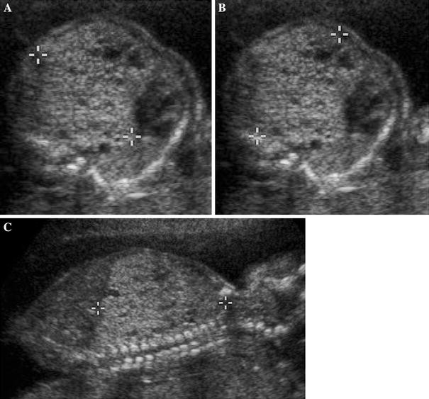
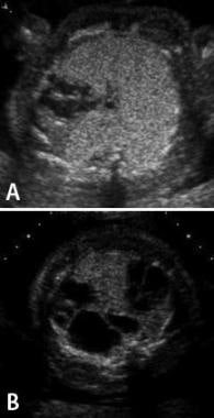
Congenital cystic lung disease: contemporary antenatal and ... from media.springernature.com Lung imaging settings for specific ultrasound systems. Much of lung ultrasound is based on the interpretation of artefacts produced at the pleural surface. The widespread use of antenatal ultrasound examination has resulted in an increase in the prenatal diagnosis of cpam 3,4. It is used to create an image of internal body structures such as tendons, muscles, joints, blood vessels, and internal organs. Fetuses that get sick exhibit hydrops fetalis on ultrasound. Lung ultrasound is increasingly used in critically ill patients as an alternative to bedside chest radiography, but the best training method remains uncertain. Congenital pulmonary airway malformations (cpam) are multicystic masses of segmental lung tissue with abnormal bronchial proliferation. Lung us is a comparatively recent addition to emergency us.
Although rare, it is the most common congenital lung lesion.
Hyperechoic, well defined, no ↓ intensity with depth. Learn lung ultrasound with expert instructor dr. Only in the last decade, it has been shown that the us assessment of the lung could have a role in common clinical practice 8. Alternative method in intensive care unit and emergency department 8. It concluded that lung ultrasound could help the clinician make a rapid diagnosis in patients with acute respiratory failure. Fast and accurate diagnosis at the bedside for clinicians, rts, and medical students from a variety of medical specialties. Cpams are considered part of the spectrum of bronchopulmonary foregut malformations. Since then, critical care doctors and respiratory physicians worldwide have taken it up for a number of indications including Spleen left lower border of lung. Right lower border of lung. We aimed to describe lu findings in cpam neonates needing. Lung ultrasound imaging may assist in the efficient allocation of intensive care for patients with respiratory failure from viral pulmonary infections, especially in resource scarce settings or situations such as future respiratory virus outbreaks or pandemics. Congenital pulmonary airway malformation (cpam) is a developmental malformation of the lower respiratory tract.
Fetuses that get sick exhibit hydrops fetalis on ultrasound. After reading this lung ultrasound tutorial you. We aimed to describe lu findings in cpam neonates needing neonatal intensive care unit (nicu). It concluded that lung ultrasound could help the clinician make a rapid diagnosis in patients with acute respiratory failure. Right lower border of lung.
Fetal Surgery for Congenital Pulmonary Airway Malformation ... from img.medscapestatic.com A simple ultrasound analysis of lungs (and veins in suitable cases) allows to categorize the test in one of seven characteristic profiles. Since then, critical care doctors and respiratory physicians worldwide have taken it up for a number of indications including Fluid around the heart, lungs, intestine as well as thickening of the skin or placenta, and. Right lower border of lung. Ultrasound can be used to explore most of the pleural surface, with the exception of the mediastinal reflections. Despite the difficulties in imaging an organ largely filled with air, the potential benefits originating from an effective ultrasound method focusing on monitoring and diagnosing lung diseases represent a. Lungs are traditionally considered poorly accessible to ultrasound (us) investigation because of their air content 7. Learn lung ultrasound with expert instructor dr.
Ultrasound can be used to explore most of the pleural surface, with the exception of the mediastinal reflections.
In the diagnosis of lung pathologies, we often use ultrasound artifacts, arising from the chest wall and pleural surface, as an interpretation. Only in the last decade, it has been shown that the us assessment of the lung could have a role in common clinical practice 8. Lung us is a comparatively recent addition to emergency us. Spleen left lower border of lung. This program introduces and defines basic lung ultrasound signs. Congenital pulmonary airway malformations (cpam) are multicystic masses of segmental lung tissue with abnormal bronchial proliferation. Congenital pulmonary airway malformation (cpam) is a developmental malformation of the lower respiratory tract. We aimed to describe lu findings in cpam neonates needing neonatal intensive care unit (nicu). Lung ultrasound is increasingly used in critically ill patients as an alternative to bedside chest radiography, but the best training method remains uncertain. Fetuses that get sick exhibit hydrops fetalis on ultrasound. Cpam (earlier ccam), pulmonary sequestration and other conditions are discussed in detail. It requires the mastery of ten signs: Enjoy a series of concise videos and case studies as you move towards mastery of point of care lung ultrasound.
This is an ideal tutorial for students & consltants practising fetal medicine to understand fetal echogenic lungs in detail. It is used to create an image of internal body structures such as tendons, muscles, joints, blood vessels, and internal organs. Lung ultrasound is a basic application of critical ultrasound, defined as a loop associating urgent diagnoses with immediate therapeutic decisions. Hyperechoic, well defined, no ↓ intensity with depth. Lungs are traditionally considered poorly accessible to ultrasound (us) investigation because of their air content 7.
Congenital Pulmonary Airway Malformation | Radiology Key from radiologykey.com Congenital pulmonary airway malformations (cpam) are multicystic masses of segmental lung tissue with abnormal bronchial proliferation. Congenital pulmonary airway malformation (cpam) is a developmental malformation of the lower respiratory tract. Hyperechoic, well defined, no ↓ intensity with depth. Its systematic use was first described by lichtenstein in his textbook 'general ultrasound in the critically ill' (springer, 2002). Lung ultrasound is a basic application of critical ultrasound, defined as a loop associating urgent diagnoses with immediate therapeutic decisions. Lung ultrasound is increasingly used in critically ill patients as an alternative to bedside chest radiography, but the best training method remains uncertain. International guidelines for lung ultrasound key article: It is used to create an image of internal body structures such as tendons, muscles, joints, blood vessels, and internal organs.
Lung ultrasound is a standardized f i e l d , w h i c h c a n b e understood perfectly by reading static images instead of mobile ones.
Congenital pulmonary airway malformation (cpam), formerly known as congenital cystic adenomatoid malformation (ccam), is a congenital disorder of the lung similar to bronchopulmonary sequestration. Lung us is a comparatively recent addition to emergency us. Lung ultrasound (lus) is a growing and fascinating field of application for ultrasound imaging. Spleen left lower border of lung. Learn lung ultrasound with expert instructor dr. Lungs are traditionally considered poorly accessible to ultrasound (us) investigation because of their air content 7. Despite the difficulties in imaging an organ largely filled with air, the potential benefits originating from an effective ultrasound method focusing on monitoring and diagnosing lung diseases represent a. Lung ultrasound imaging may assist in the efficient allocation of intensive care for patients with respiratory failure from viral pulmonary infections, especially in resource scarce settings or situations such as future respiratory virus outbreaks or pandemics. Lung imaging settings for specific ultrasound systems. This program introduces and defines basic lung ultrasound signs. In the diagnosis of lung pathologies, we often use ultrasound artifacts, arising from the chest wall and pleural surface, as an interpretation. Fetuses that get sick exhibit hydrops fetalis on ultrasound. Lung ultrasound is a basic application of critical ultrasound, defined as a loop associating urgent diagnoses with immediate therapeutic decisions.
A simple ultrasound analysis of lungs (and veins in suitable cases) allows to categorize the test in one of seven characteristic profiles cpam. Fetuses that get sick exhibit hydrops fetalis on ultrasound.


0 Komentar